.jpg) As the lead design engineer at Kelyniam Global, I am constantly faced with new challenges. Each case is unique as are the needs for the patients receiving implants. Much has changed since the first cranial and facial implants were developed, and exponentially so in the last decade.
As the lead design engineer at Kelyniam Global, I am constantly faced with new challenges. Each case is unique as are the needs for the patients receiving implants. Much has changed since the first cranial and facial implants were developed, and exponentially so in the last decade.
New materials – and improved design and manufacturing techniques – mean much better outcomes for patients in form, fit, and function. In addition, increased computer power and greatly improved software have pushed the industry like never before.
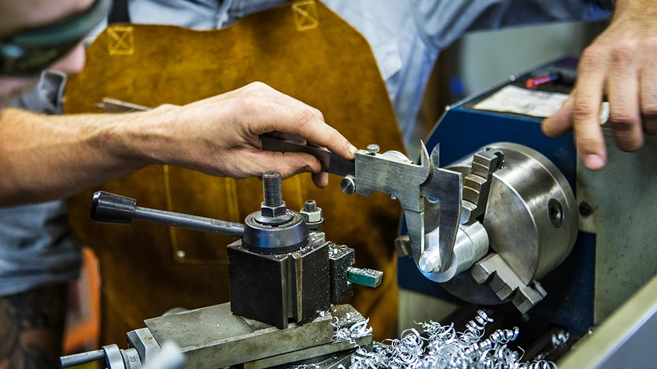
Significant advancements in the use of Bio-CAD/CAM technology now allow us to provide near-perfectly replicated cranial and craniofacial implants to replace damaged bone structures. Derived from the patient’s CT scan data, 3D BIO-CAD/CAM software transfers the intricate details found on the edge of the defect directly to the implant edge. Computer numerical control (CNC) technology precision mills the implants – using tolerances that are less than 0.010", resulting in an exact replication of the patient’s missing bone flap.
Design challenges
I was recently tasked with designing a rigid, passive implant for a highly complex craniofacial case. We received the customer-supplied CT data of the defective area via the Internet. This data were then converted into a workable 3D digital model that could be used in a CAD application. The model showed massive reconstruction of the facial bones as well as trauma in the left facial region caused by a gunshot wound.
Initially, I prepared a one-piece implant, as is customary, but the surgeon realized implanting the device in its current state would require a large incision – never an optimal thing in facial surgery – and requested a redesign. After considerable thought, I came back with a four-piece part that would fit through a much smaller incision, could be assembled during implantation, and would reduce scarring and swelling, providing a better structural and aesthetic outcome.
The unique four-part design mimics the form and function of healthy bone while reducing surgery and patient-recovery time. We achieved this by greatly minimizing the entry incision and aiding the surgeon in part placement and assembly. This also combined to reduce the risk of infection.
There were a number of significant challenges to overcome when designing and machining the four complementary parts. The treatment area was complex, with rough, broken facial bones and hardware from a previous surgery. Part placement had the potential to be problematic, since most of the orbital floor was missing, creating a risk that the implant could drop out of position before it could be secured. Furthermore, fastening the four parts together without sacrificing part integrity or increasing the risk of infection or irritation for the patient created its own set of complications.
|
About Kelyniam Kelyniam specializes in the production of custom prosthetics using computer-aided design and computer-aided manufacturing of advanced medical-grade polymers. As a producer of custom polyether ether ketone (PEEK) cranial and facial implants, Kelyniam can design and manufacture implants in as little as 24 hours from receipt of order. Reducing the gap between trauma and implantation benefits surgeons and patients – as well as health care budgets – by shortening hospital stays. The precise anatomic fit reduces operating time compared to traditional reconstructive methods, such as titanium mesh/bone cement, that require extensive contouring. |
The implant’s orbital floor was about half the size of a normal one. This aided in part placement through the smaller-than-normal incision. The inferior orbital rim implant was built out with a supporting on-lay lip to hold the orbital floor implant. The nasal bone and frontal process of the maxilla implant were built up for aesthetic reasons and were joined against the orbital floor implant in a two-dimensional ball-and-socket joint. This design allowed the surgeon to drop in the part near its final location and slide it into place prior to fastening.
Manufacturing challenges
To reduce the risk of infection or irritation, the part edges were rounded and smoothed. Our manufacturing division used proprietary CNC machining techniques to manufacture the multifaceted parts. The parts were designed with a precise surface mirroring on-lay technique over each surface imperfection to achieve optimal part placement. This meant the small intricate plastic parts would have to be manufactured with new techniques. Since the parts were precisely manufactured, this provided a unique identifier on the patient’s bone to allow for a custom drop-in fit that could hold the implant securely in place while the surgeon placed the fasteners. In other words, each implant would on-lay exactly on top of the bone’s surface. This unique identifier on-lay technique was used to help reduce lateral movements and improve part placement during surgery. We believe this type of design and procedure will become much more commonplace.
Material choice
We work closely with a trusted materials partner in the medical plastics market, Invibio. Invibio transformed the medical device design landscape with the introduction of its PEEK-OPTIMA polymer family 15 years ago and set an industry standard for biomaterials biocompatibility in safety and quality. PEEK-OPTIMA Polymer’s proven biocompatibility is supported by U.S. FDA and China CFDA Drug & Device Master Files, and has been used in more than 4 million approved, implantable devices worldwide. These strengths have encouraged the orthopedic industry to use PEEK-OPTIMA materials in many other applications, including hip, knee, and finger joints, plus arthroscopy anchors and screws.
Kelyniam Global
www.kelyniam.com
About the author: Merwin Schaefer is the lead design engineer for Kelyniam Global and can be reached at 800.280.8192 or info@kelyniam.com.

Explore the March 2015 Issue
Check out more from this issue and find your next story to read.
Latest from Today's Medical Developments
- NextDent 300 MultiJet printer delivers a “Coming of Age for Digital Dentistry” at Evolution Dental Solutions
- Get recognized for bringing manufacturing back to North America
- Adaptive Coolant Flow improves energy efficiency
- VOLTAS opens coworking space for medical device manufacturers
- MEMS accelerometer for medical implants, wearables
- The compact, complex capabilities of photochemical etching
- Moticont introduces compact, linear voice coil motor
- Manufacturing technology orders reach record high in December 2025