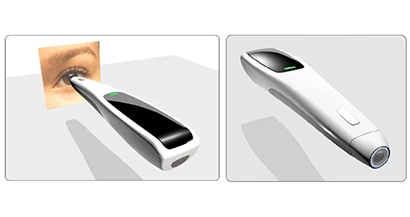
Last week, Congresswoman Louise Slaughter (NY-25) visited Rochester, N.Y.-based medical device technology firm Lumetrics to announce a $973,000 research and development grant from the National Institutes of Health – National Eye Institute. The NIH grant will fund development of a digital hand-held diagnostic ophthalmic instrument that will dramatically improve access to vision related healthcare both in the U.S. and across the globe. The promising pen-sized device will provide an effective clinical tool for inspecting the human retina and documenting the findings.
“The benefits that this innovation will provide both here in the U.S. and across the globe are immeasurable. The ability to provide comprehensive diagnostic eye care with a hand-held device is truly groundbreaking and represents a huge step forward in improved healthcare access for previously underserved populations.” Slaughter, a degreed microbiologist, says.
Vision is one of the most valued human sensory experiences. Vision loss can be caused by many factors, stemming from damage to all parts of the visual system. Retinal and optic nerve problems have emerged as leading causes of visual loss in developed countries. Fortunately, many of these conditions are treatable. In most cases, early diagnosis and proper follow up lead to adequate maintenance of visual function for life. Furthermore, the early diagnosis of conditions such as dry age-related macular degeneration can help patients address risk factors for progression, thereby delaying and possibly preventing long-term visual loss.
Lumetrics, a 20-person measurement systems company founded in 2003, has developed innovative measurement systems. The company currently counts among its customers six of the top 11 medical device manufacturers in the world. This latest development is an extension to a previous grant that demonstrated the feasibility of this technology. This new grant is for the miniaturization and continuation of that project.
“We are extremely pleased that the National Institutes of Health found our idea worthy of such a large and prestigious grant,” states Lumetrics’ CEO John Hart. “The collaboration with the University of Rochester and its Flaum Eye Institute is an incredible opportunity for Lumetrics and will lead to new treatments in vision care.”
The plan for commercialization is centered on the idea that the proposed device represents a 21st century replacement for the direct ophthalmoscope. The direct ophthalmoscope in its basic configuration has been the routine method of retinal examination for the past 100 years. The market analysis and the interviews conducted with physicians, specialists, and technicians shows significant need for this new, more sophisticated device.
This digital ophthalmoscope will be used by general practitioners, family practice physicians, internists, and pediatricians during patient visits, and lead to meaningful accurate documentation of retinal disease status, and follow the disease progression over a period of time. This device will drastically improve patients’ access to the high quality fundus images required to manage retinal and optic nerve diseases on the early stage. The device’s success will be felt throughout the world especially where the lack of access to fundus examination is severe.
The development of the new camera is being led by Filipp Ignatovich, PhD, Chief Technology Officer for Lumetrics and David Kleinman, MD, MBA an academic retinal specialist at the Flaum Eye Institute.
Dr. Filipp Ignatovich is an expert in developing innovative strategies to leverage technology in improving the human condition. He defines and oversees the research and development efforts at Lumetrics with the emphasis on medical ophthalmic devices.
“I am fortunate to lead a great team of professionals in developing this instrument which has the potential to make a positive impact in health and vision care for all people,” Ignatovich says. “This funding exemplifies meaningful government initiatives designed to promote innovation, small business success, and cost effective interventions advancing quality of life.”
The functionality of the camera is simplified and automated to minimize the demand on the operator skill. The technology behind the camera is made possible through a key patent invented by Steven Feldon, MD, MBA, Director of the Flaum Eye Institute and Geunyoung Yoon, PhD, of the University of Rochester, both of whom will be assisting on aspects of the project.
The previous phase of the project provided Lumetrics with a good understanding of the technical challenges and pitfalls associated with imaging and illumination of the retina of the eye. The following challenges must be conquered as part of this grant.
- Reducing the minimum pupil size
- Maintaining uniform illumination while obtaining proper image
- Obtaining and automating acceptable touch pressure on the eye
- Determining proper form factor for the device and components
- Obtaining correct color mapping, image analysis and feature recognition of the eye
The Phase I development project met its goals; Lumetrics developed a proof-of-principle prototype of the ultra-compact fundus camera, and clinically proved its acceptable performance. This experience and knowledge will allow for effective and speedy development during this new stage of the project.
Latest from Today's Medical Developments
- NextDent 300 MultiJet printer delivers a “Coming of Age for Digital Dentistry” at Evolution Dental Solutions
- Get recognized for bringing manufacturing back to North America
- Adaptive Coolant Flow improves energy efficiency
- VOLTAS opens coworking space for medical device manufacturers
- MEMS accelerometer for medical implants, wearables
- The compact, complex capabilities of photochemical etching
- Moticont introduces compact, linear voice coil motor
- Manufacturing technology orders reach record high in December 2025