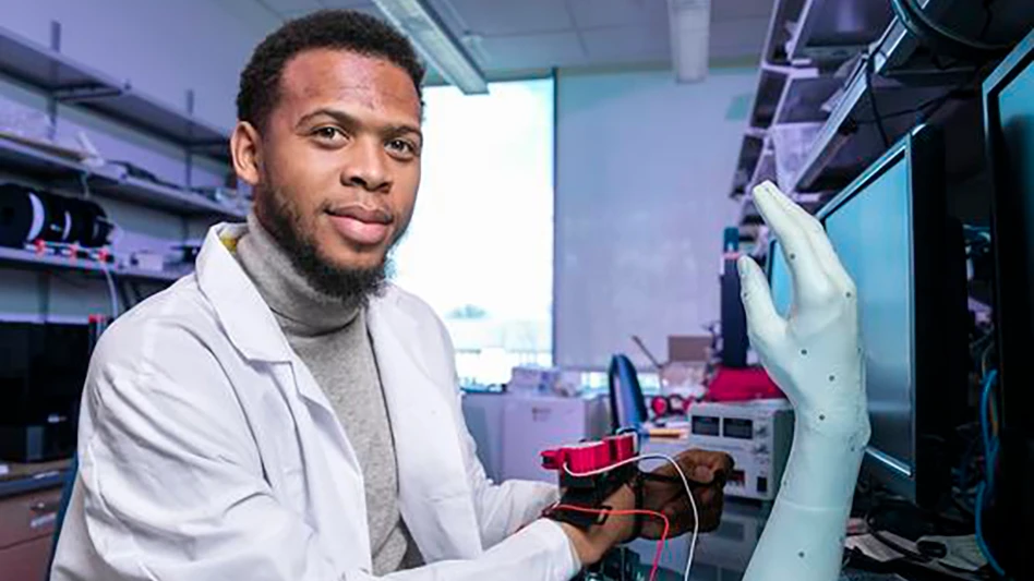
Modern tissue engineering developed at the University of Michigan Health System could improve the function of prosthetic hands and possibly restore the sense of touch for injured patients.
Researchers will present their updated findings Wednesday at the 95th annual Clinical Congress of the American College of Surgeons. The research project, funded by the Department of Defense, arose from a need for better prosthetic devices for troops wounded in Afghanistan and Iraq.
“Most of these individuals are typically using a prosthesis design that was developed decades ago,” says Paul S. Cederna, M.D., a plastic and reconstructive surgeon at U-M Health System and associate professor of surgery at the U-M Medical School. “This effort is to make a prosthesis that moves like a normal hand.”
U-M researchers may help overcome some of the shortcomings of existing robotic prosthetics, which have limited motor control, provide no sensory feedback and can be uncomfortable and cumbersome to wear.
“There is a huge need for a better nerve interface to control the upper extremity prostheses,” Cederna says.
When a hand is amputated, the nerve endings in the arm continue to sprout branches, growing a mass of nerve fibers that send flawed signals back to the brain.
The researchers created what they called an “artificial neuromuscular junction” composed of muscle cells and a nano-sized polymer placed on a biological scaffold. Neuromuscular junctions are the body's own nerve-muscle connections that enable the brain to control muscle movement.
That bioengineered scaffold was placed over the severed nerve endings like a sleeve.
The muscle cells on the scaffold and in the body bonded and the body’s native nerve sprouts fed electrical impulses into the tissue, creating a stable nerve-muscle connection.
In laboratory rats, the bioengineered interface relayed both motor and sensory electrical impulses and created a target for the nerve endings to grow properly.
“The polymer has the ability to pick up signals coming out of the nerve, and the nerve does not grow an abnormal mass of nerve fibers,” Cederna explains.
The animal studies indicate the interface may not only improve fine motor control of prostheses, but can also relay sensory perceptions such as touch and temperature back to the brain.
Laboratory rats with the interface responded to tickling of feet with appropriate motor signals to move the limb, Cederna says.
The Department of Defense and the Army have already provided $4.5 million in grants to support the research. Meanwhile, the research team has submitted a proposal to the Defense Advance Research Project Agency to begin testing the bioengineered interface in humans in three years.
Additional U-M authors of the study include William M. Kuzon, Jr., M.D., Ph.D., professor of surgery and head of the Division of Plastic Surgery; David C. Martin, Ph.D., chair of materials science and engineering at the University of Delaware and former professor of materials science and engineering and macromolecular science at U-M; Daryl R. Kipke, Ph.D. professor of biomedical engineering; Melanie Urbancheck, Ph.D. research assistant in the Department of Surgery; and Brent M. Egeland, M.D., surgical resident.
Resources:
U-M Department of Surgery Division of Plastic Surgery
http://surgery.med.umich.edu/plastic/
Latest from Today's Medical Developments
- Gore completes acquisition of Conformal Medical
- Medical textiles designed for cardiovascular, orthopedic, dental prosthetic applications
- Micro-precision 3D printing: Trends and breakthroughs in medical device manufacturing
- One-component, dual-cure adhesive system for medical device assembly
- #82 Manufacturing Matters - Forecasting 2026 with GIE Media's Manufacturing Group
- Flexing prosthetic finger offers lifelike appearance and movement
- How the fast-evolving defense market impacts suppliers
- Medtronic’s Hugo robotic-assisted surgery system makes US debut